Our laboratory has developed several ex vivo models based on keeping human skin alive.
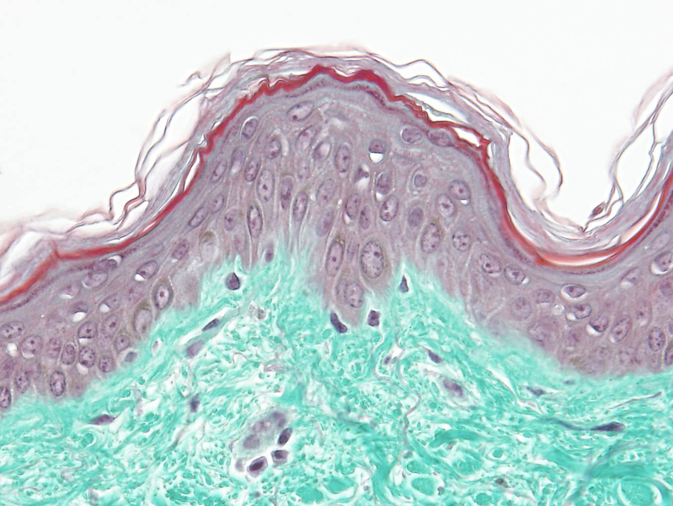
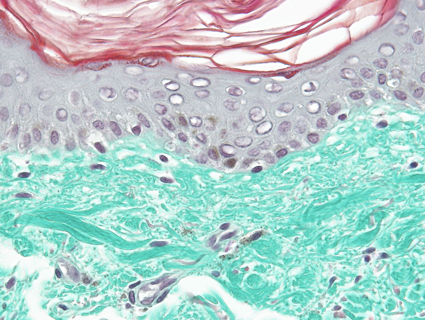
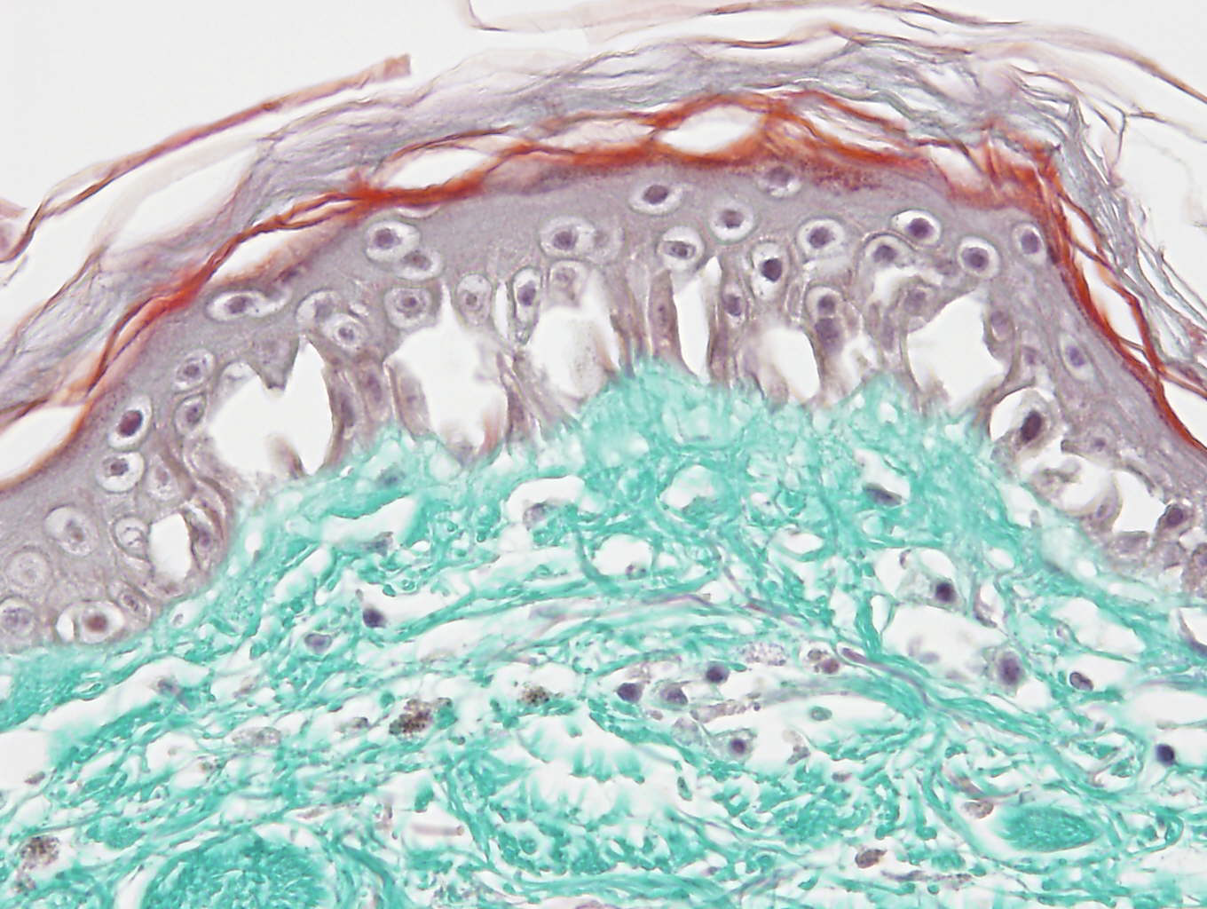
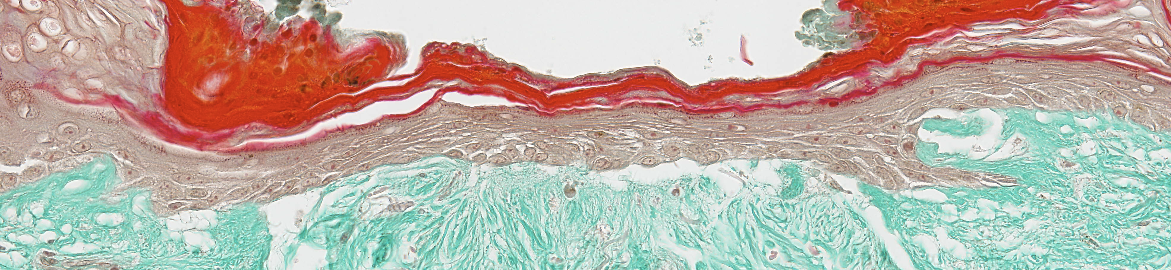
The skin conserves a normal morphology, structure and metabolism.
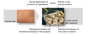
Several application modes are possible: topically or solubilised in the survival medium, single or chronic application. Possibility of making injections, laser treatments, etc.
Possibility of reproducing external interactions: exposure to UV or pollutants, chemical attacks, wound, sensitisation, hair removal, etc.
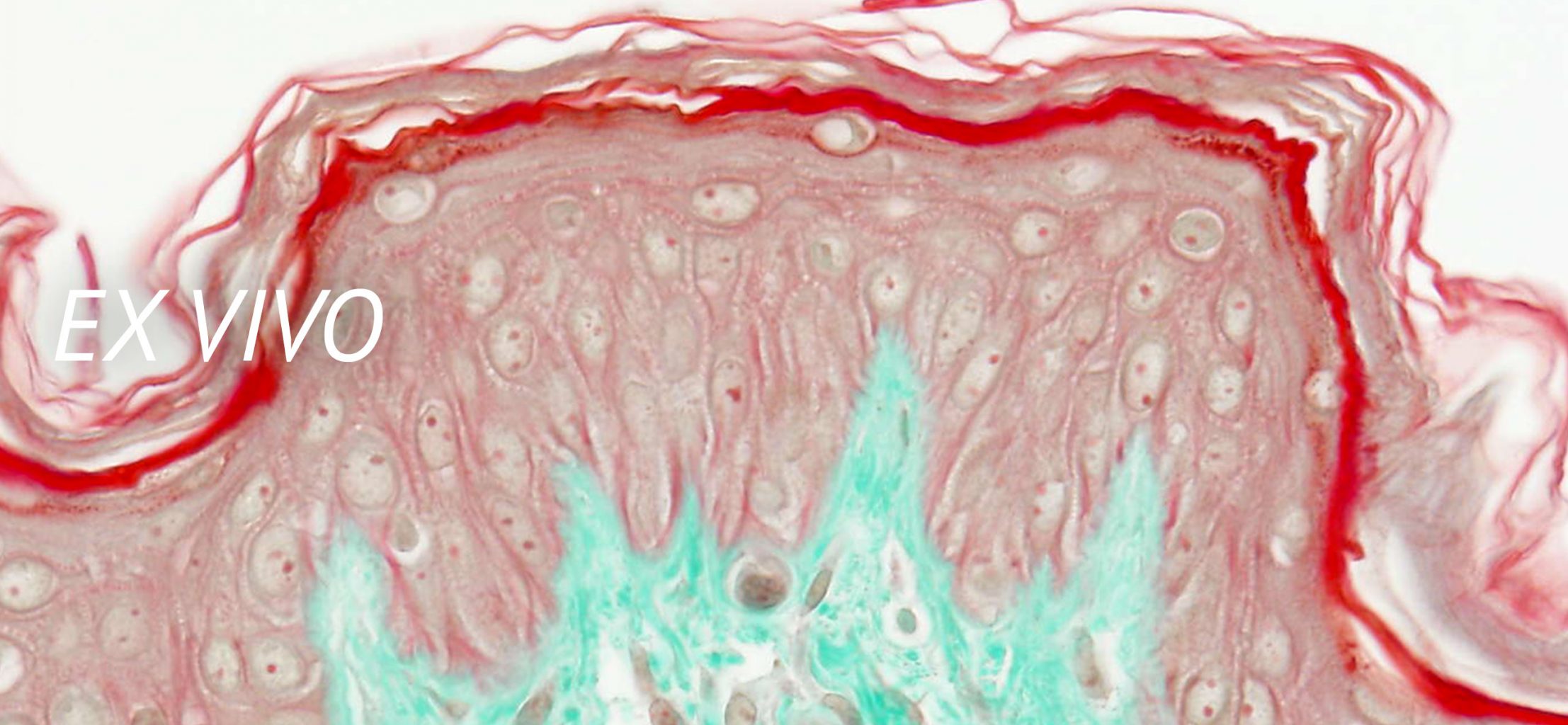
Carrying out histological analyses
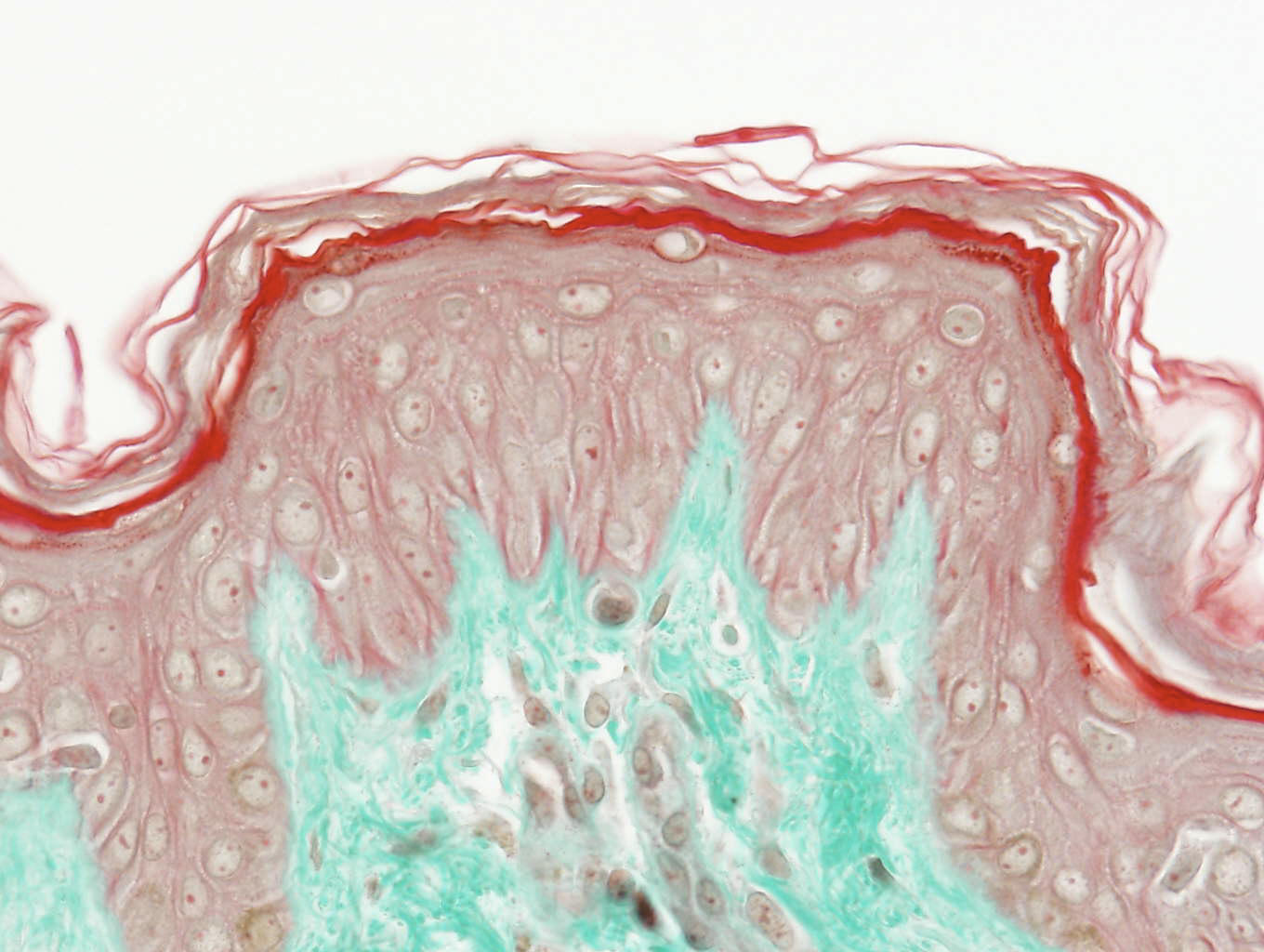
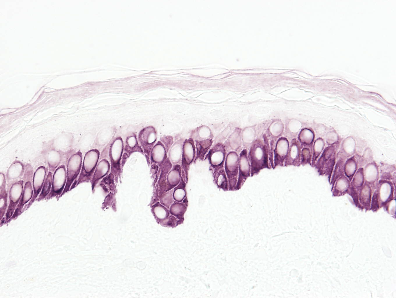
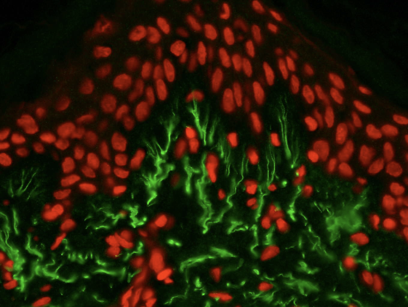
Our laboratory has a battery of stainings and immunomarkers that enable different cellular and tissue markers to be visualised. The marked parameters may be quantified by image analysis software.
Carrying out biological analyses
On whole skin, separate epidermises, homogenates, extracts or in the culture medium. All types of extractions, separations, chromatograms and assays may be envisaged.
Carrying out genomic studies (in partnership with the Genex Laboratory)
Large-scale screening and analysis of genomic data. Identification of the effects of the products on human skin thanks to the study of the genic expression (DNA chip, miRNA chip, qPCR) correlated with the protein expression of the genes of interest.
Ex vivo objectivation enables the action mechanisms of a product or active ingredient on the skin to be understood.
Furthermore, thanks to the images obtained from histological processes, our studies provide you with excellent materials for your marketing claims.
BIO-EC laboratory also develops specific, customised studies to prove your claims.
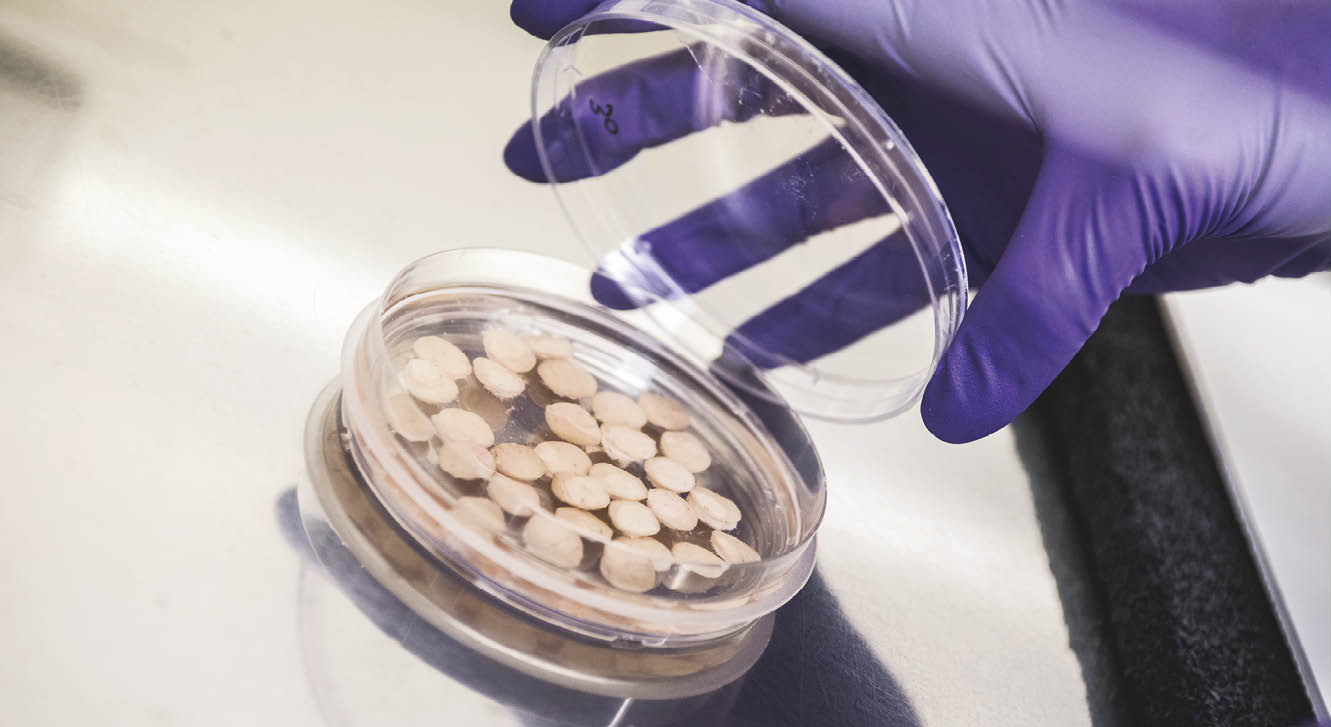
BIO-EC Laboratory has developed an ex vivo mode based on keeping explants of human skin alive for 10 to 12 days. This model enables pertinent replies to your cosmetic and dermatological objectivation requirements to be obtained.
This model enables us:
To work in conditions very close to the actual physiology of the skin, with, inter alia, all the cellular population and their communications remaining active.
To choose the phototype, the age and the gender of the donor.
To put different experimental protocols in place in order to best refine your objectivations
To rapidly observe the effects of your product on the skin ahead of in vivo studies (on volunteers).
To provide you with answers on understanding the action mechanism of your product that you can illustrate by all the photographs provided in the final study report.
Ex vivo appears to be a first-line technique, both in the diversity of the personalisable parameters for your study and in the quality of the results that you will obtain for the objectivation of your active ingredient or finished product.
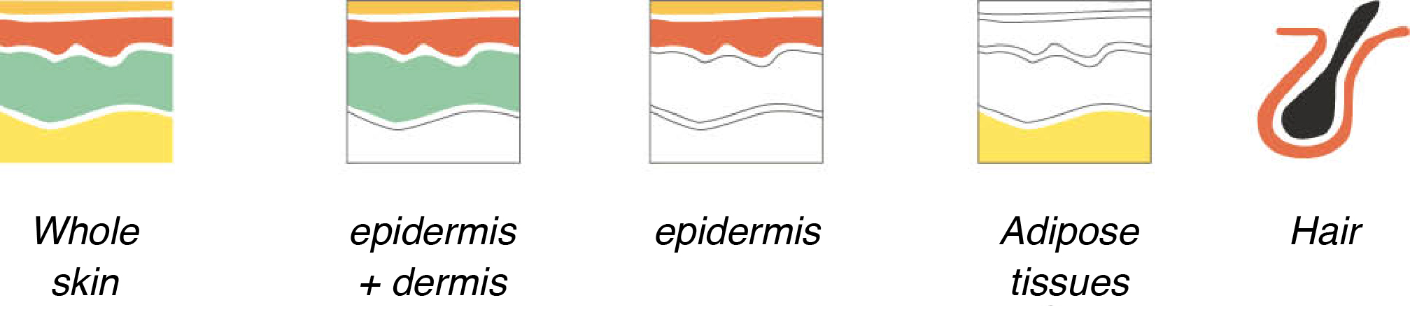
The model developed by BIO-EC Laboratory can be adapted on the basis of the products to be tested. Indeed, it is possible to carry out studies on whole skin, on isolated cutaneous compartments and on hair.
Our expertise gives us the possibility of exposing our models to different treatments and, after applying your products, different biological markers can be studied by immunomarking or assays.
All the histological studies can be accompanied by an OMICs study thanks to our collaboration with the Genex Laboratory.
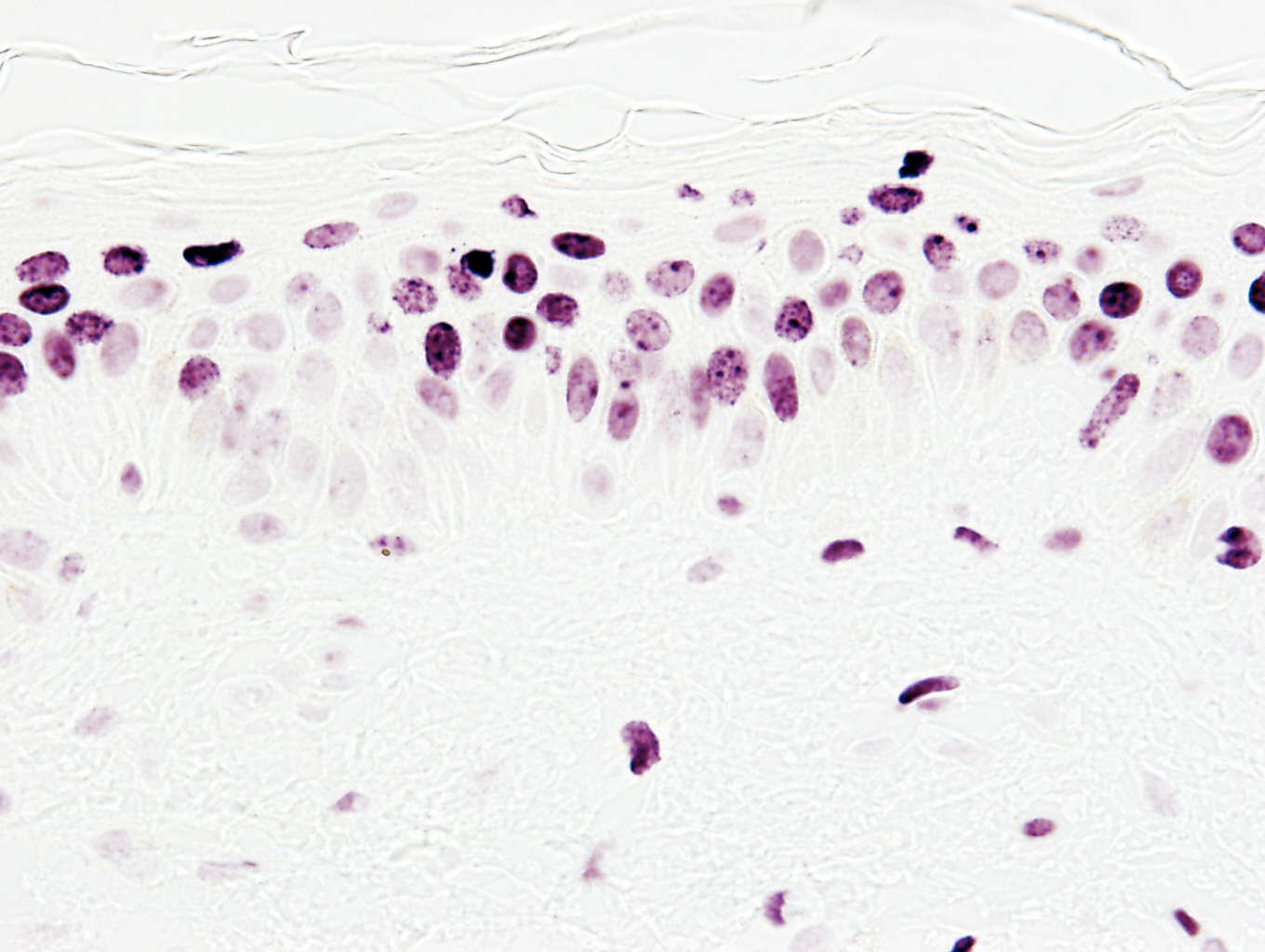
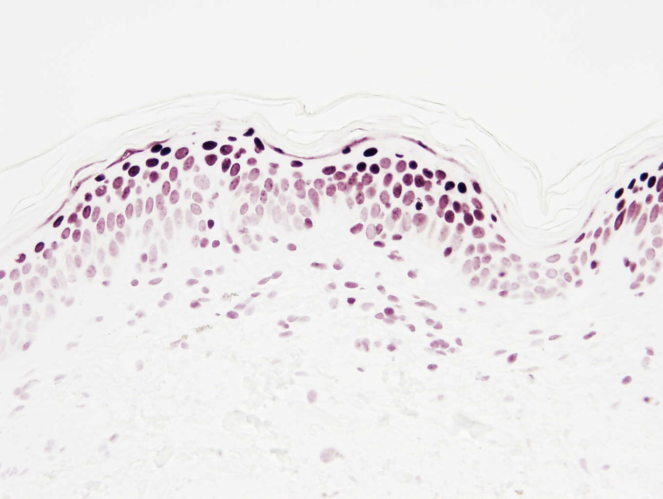
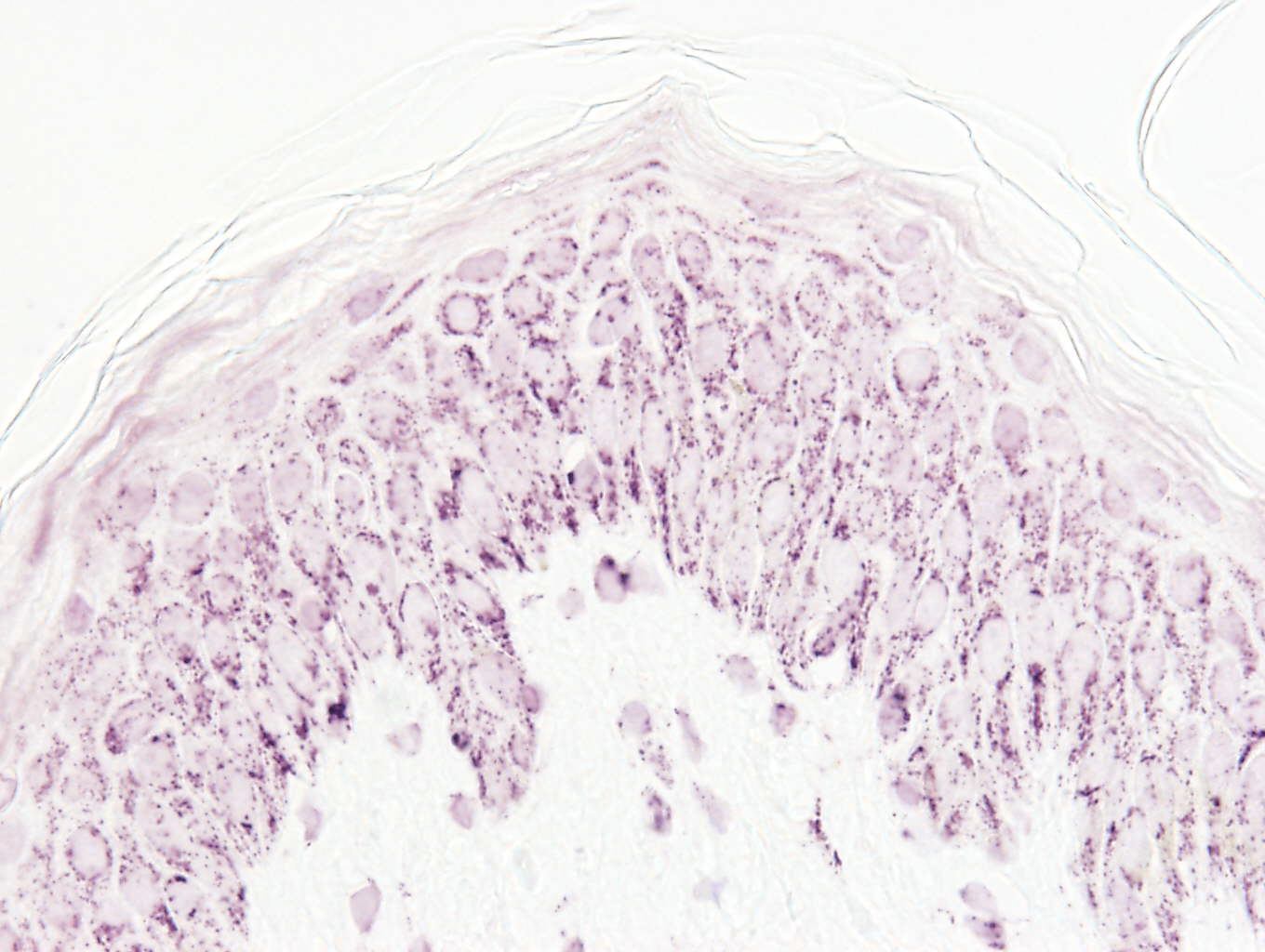
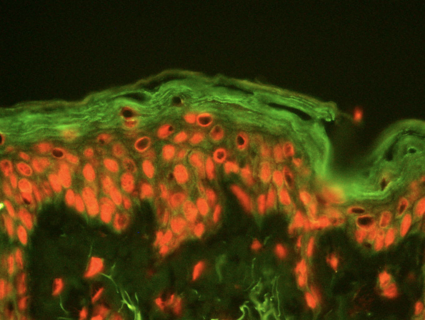
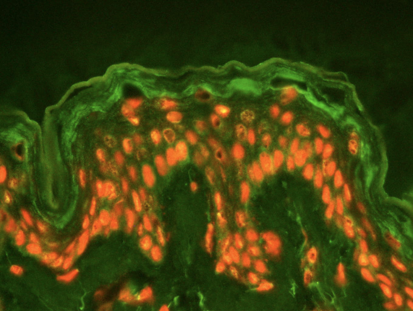
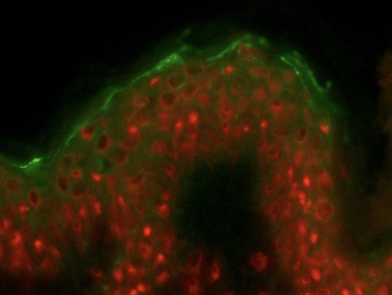
Thickness of the epidermis
Epidermal and dermal stimulation
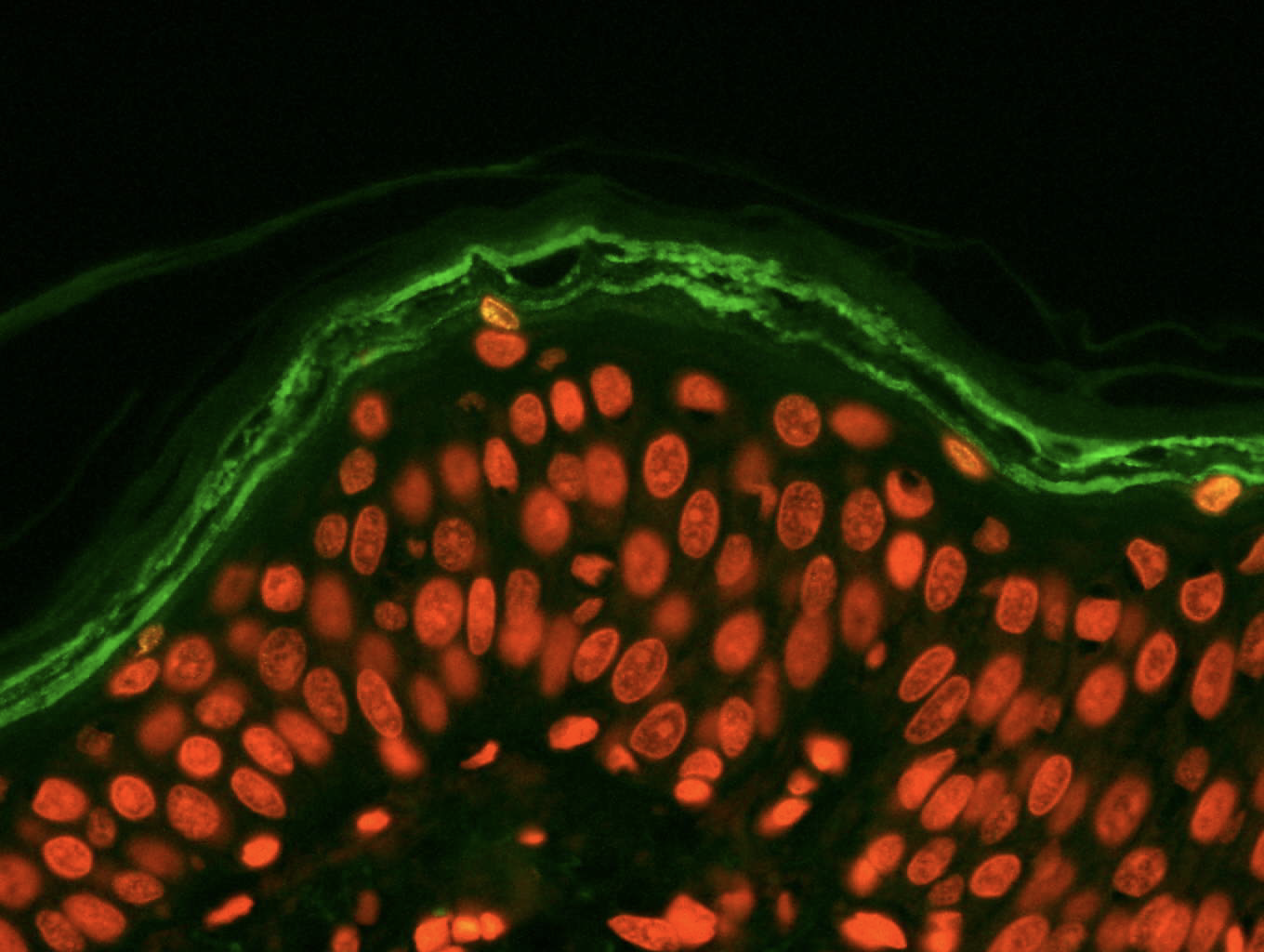
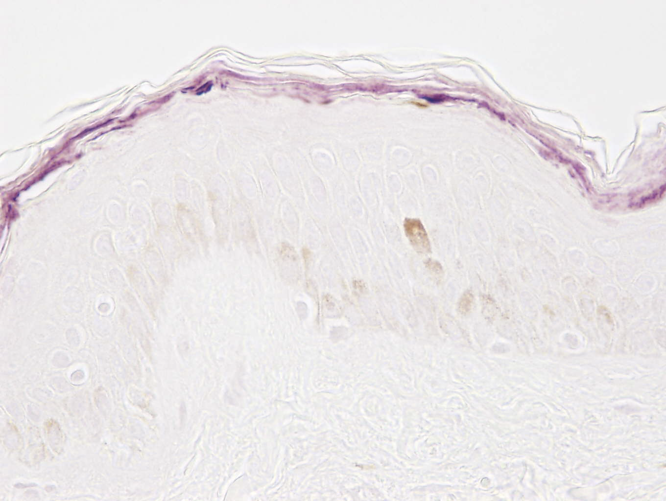
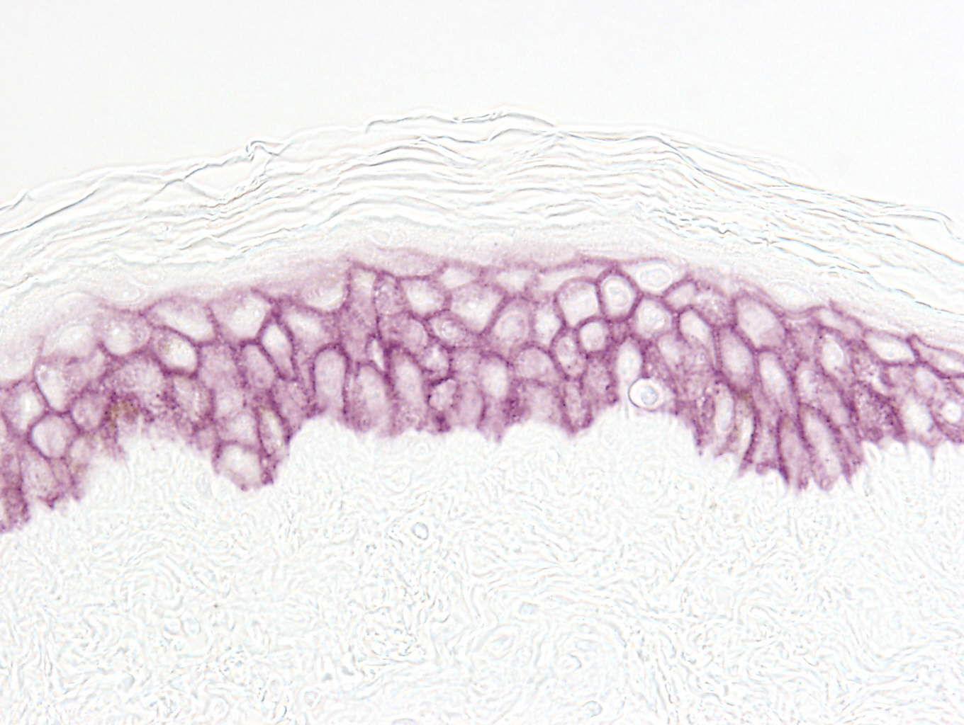
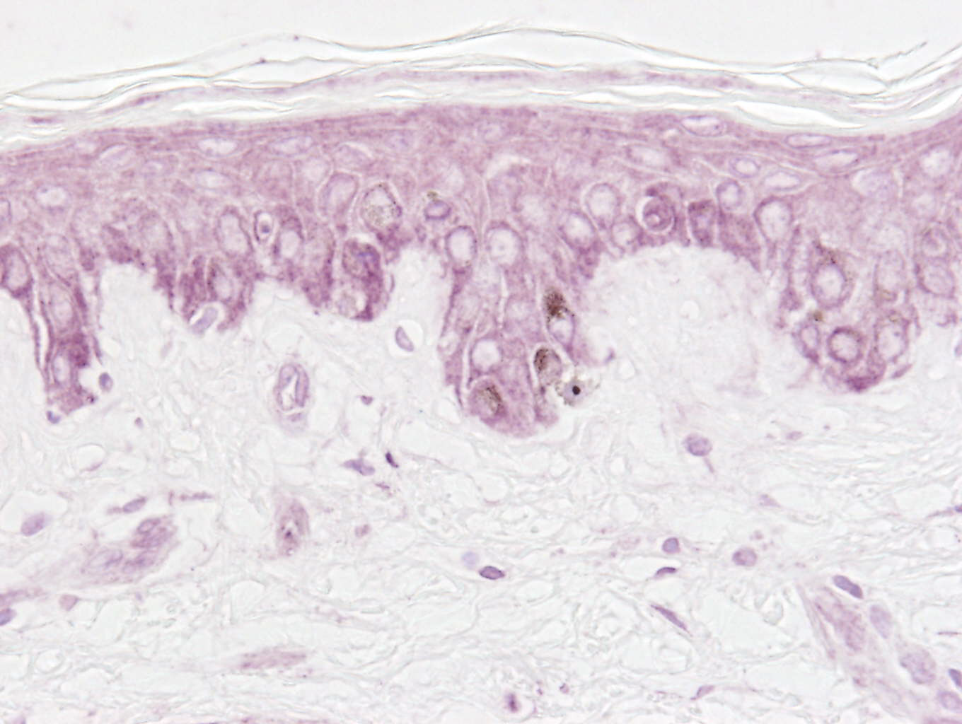
Epidermal markers
– Mitotic division (Ki67)
– Cytokeratins (CK14, CK10, CK1)
– Keratinocyte differentiation (involucrin, loricrin, caspase 14)
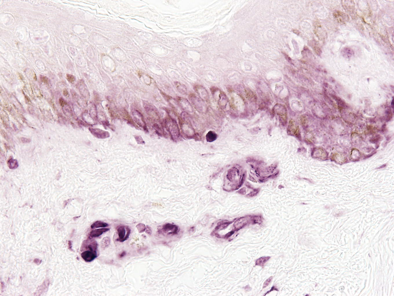
Dermal-epidermal junction markers
– Integrin B4
– Laminin 5
Dermal markers
– Extracelluar matrix (GAGs)
– Synthesis of hyaluronic acid (HAS1 and HAS2)
– Heparanase 1
– Hyaluronic acid
– Versican
– Elastin
– Fibrillin
– Emilin
– Vimentin
– Collagens (types I, III, IV, V, VII, VIII et XII)
Other markers: progerin, NDST1 and NDST2, UGT1A, etc.
With the help of this device BIO-EC Laboratory can carry out topographic studies on the surface of explants with an accuracy evaluated at a tenth of a nanometre. This enables various parameters to be analysed such as the depth and width of wrinkles and micro-lines and the roughness of the skin in order to evaluate a filling effect.
General morphology
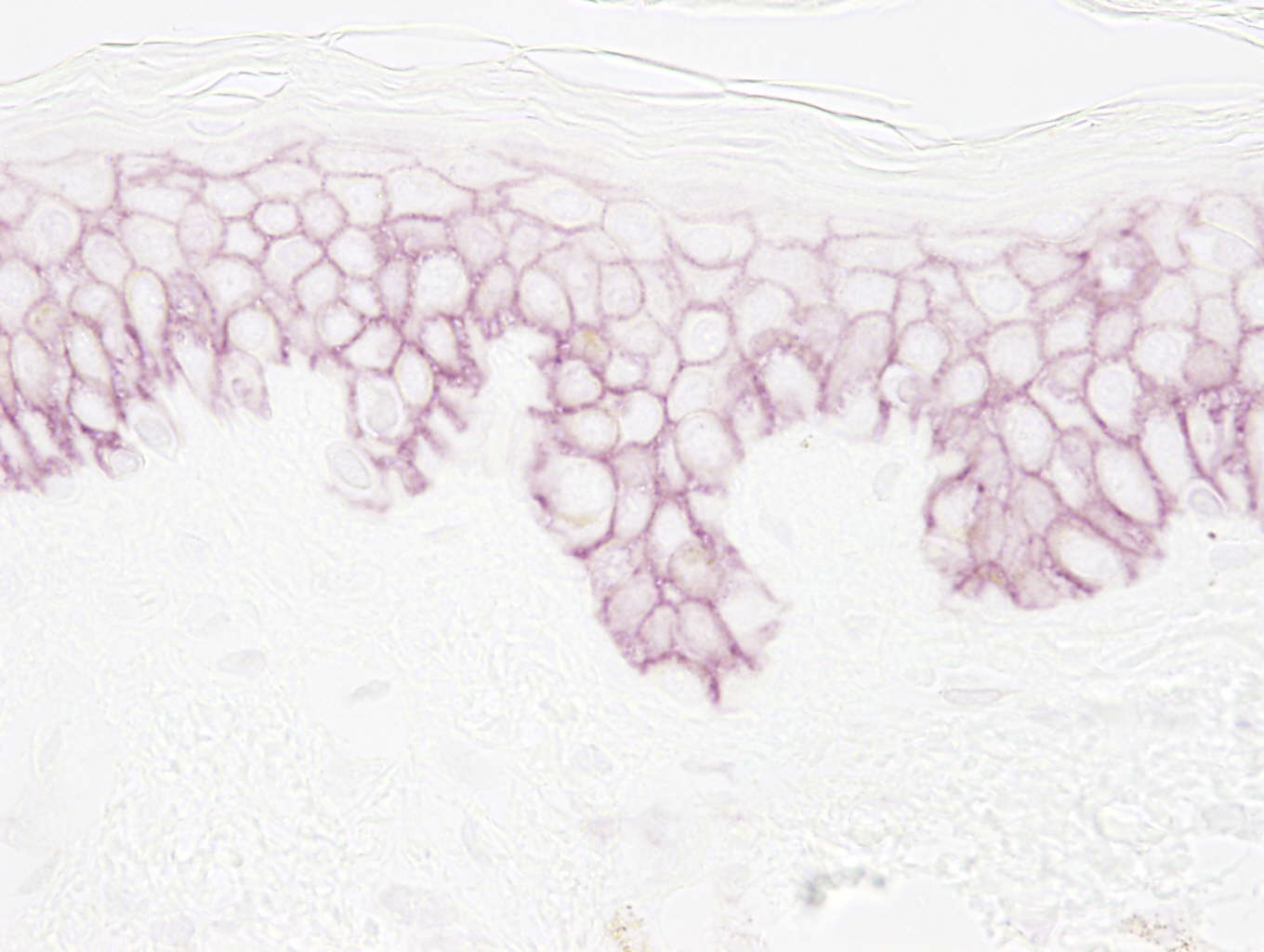
Cytokeratin 14
– ZO-1
– Occludin-1
– Claudin-1
– Loricrin
– Involucrin
– Caspase 14
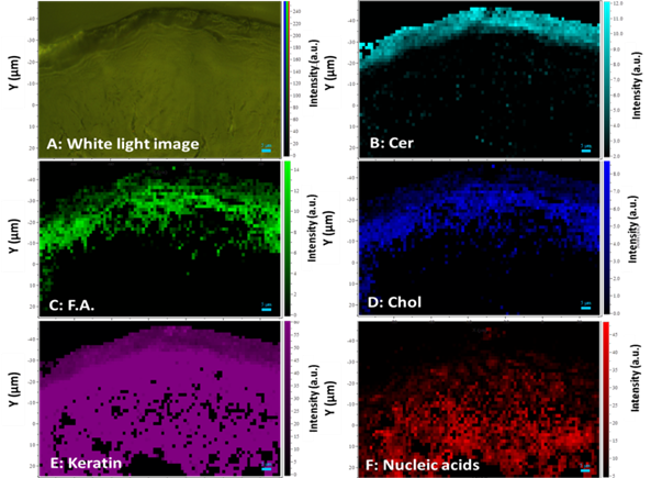
BIO-EC Laboratory may, using this device, evaluate the kinetics of the tissue penetration and distribution of your product, the skin modifications caused by your product across various parameters, the hydration rate in the stratum corneum and the epidermis, the distribution of the NMF components, of the cutaneous lipids, the carotenoids, etc.
Thanks to our partnership with BIC (Bordeaux Imaging Center), it is possible to complement our histological studies with TEM observations, such as the evaluation of the quality of lipid bilayers.
With the help of this devic BIO-EC Laboratory can carry out topographic studies on the surface of explants with an accuracy evaluated at a tenth of a nanometre. This enables various parameters to be analysed such as the depth and width of wrinkles and micro-lines and the roughness of the skin in order to evaluate a filmogenic effect.
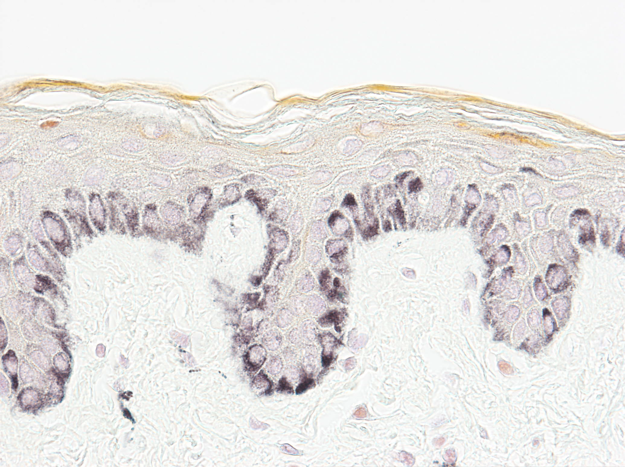
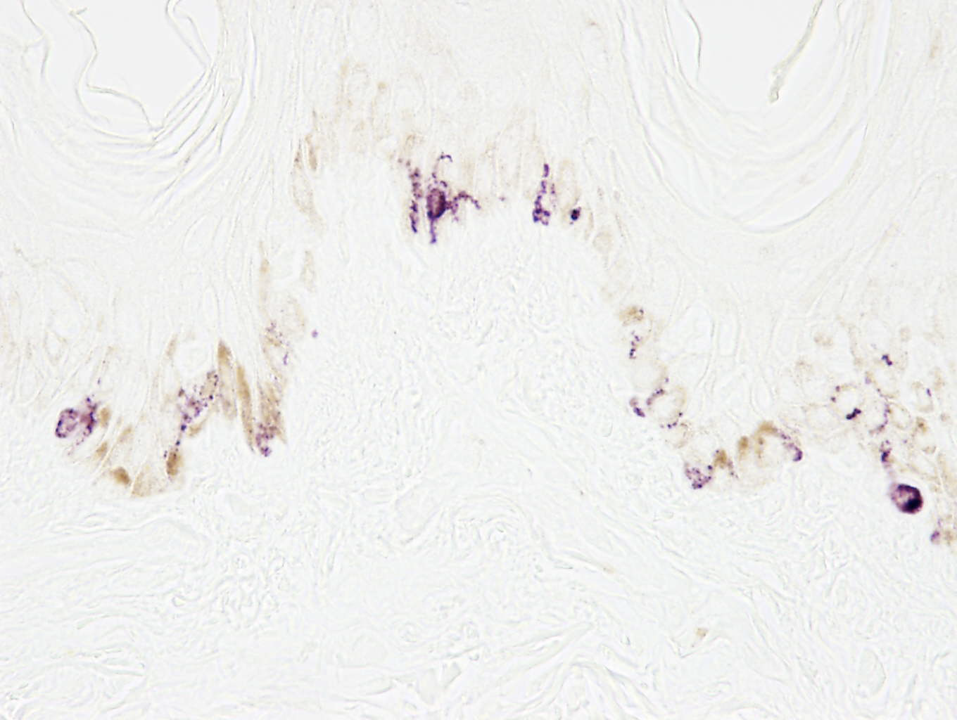
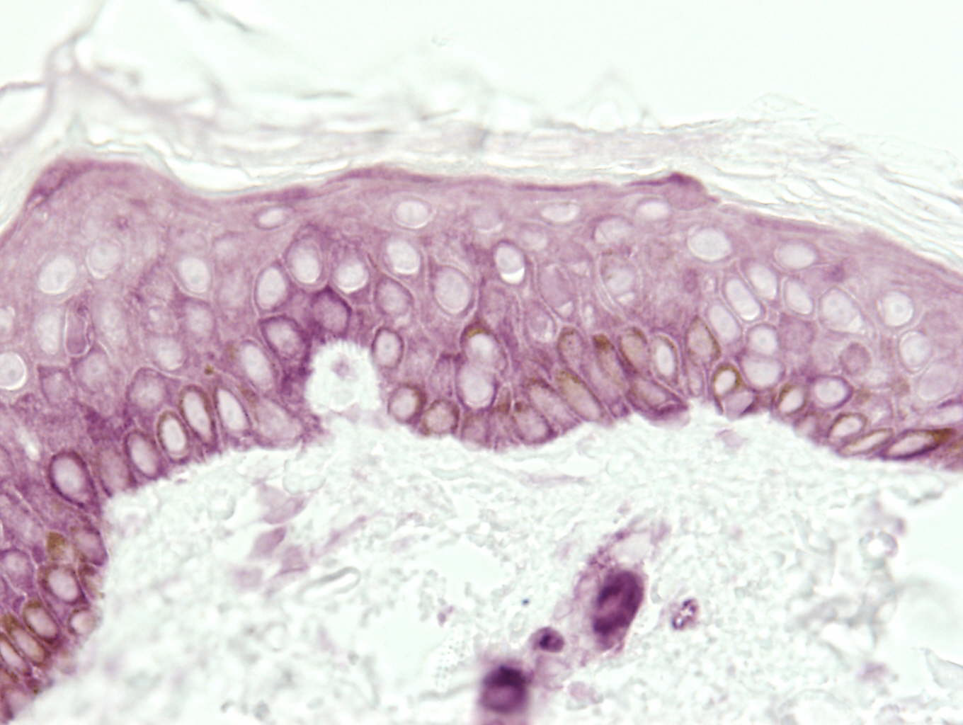
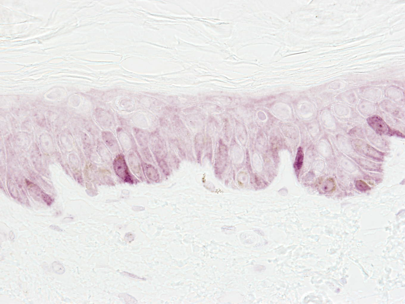
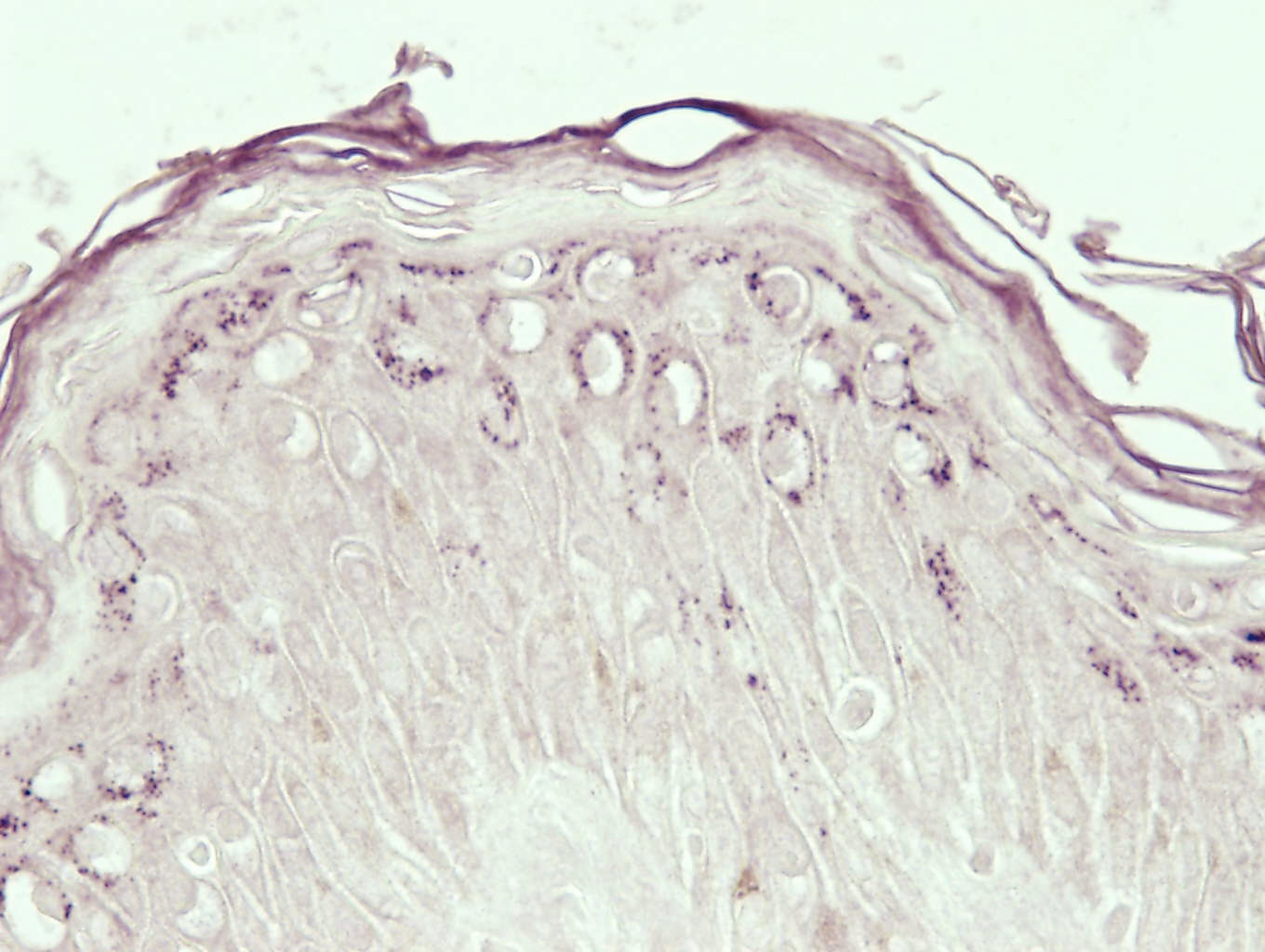
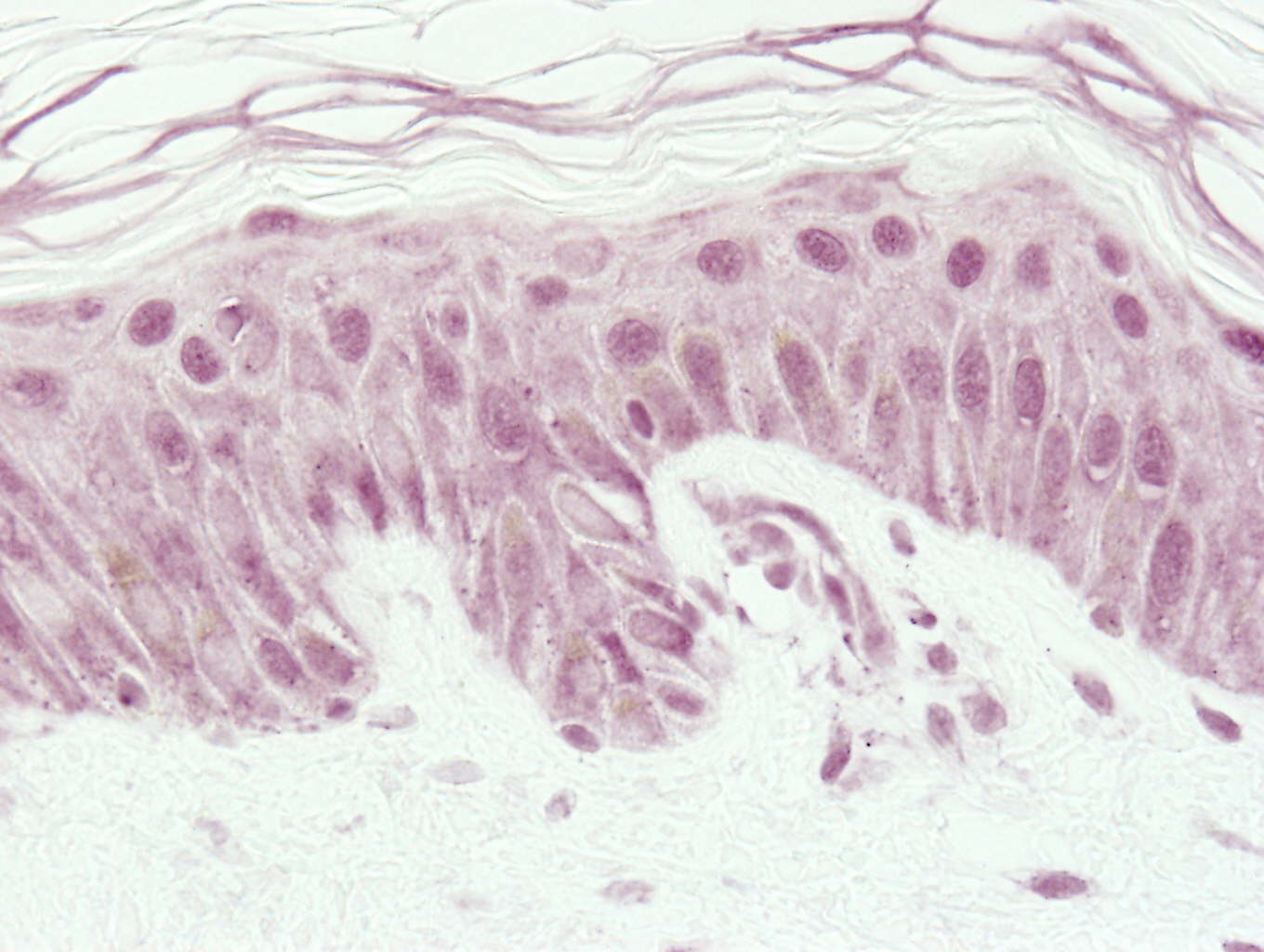
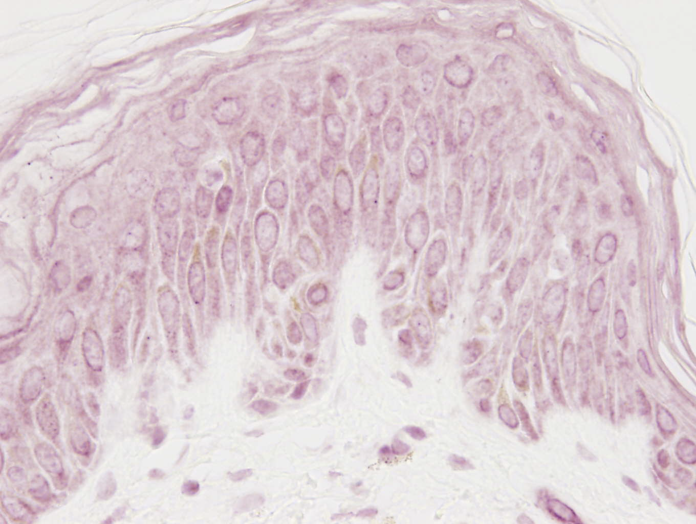
Visualisation of melanin by Fontana-Masson.

Screening test to evidence melanin produced by melanocytes: DOPA oxidase test
Test carried out on separate epidermis.
UVs (UltraViolets)
IR (infrared)
Visible light
SOLARBOX®
Process exclusive to BIO-EC Laboratory which enables us to expose the skin solely to visible light. It is also possible to expose the skin to each luminous spectrum independently:
– Full visible domain: 420-650 nm
– Band in the blue: 420-480 nm
– Band in the yellow-green: 90-650 nm
– Band in the red: 575-645 nm
Observation of the appearance of Sun Burn Cells (SBCs).
DNA protection after UVB irradiation
Evaluation of the oxidative stress after UVA irradiations
(mitochondrial enzyme)Markers evaluated after infrared ray irradiation
Markers evaluated after irradiation by visible light (blue light)
MDA (malonedialdehyde acid), lipid peroxidation marker.
Cytokines

Tropoelastin
Thymin dimers
In the culture medium:
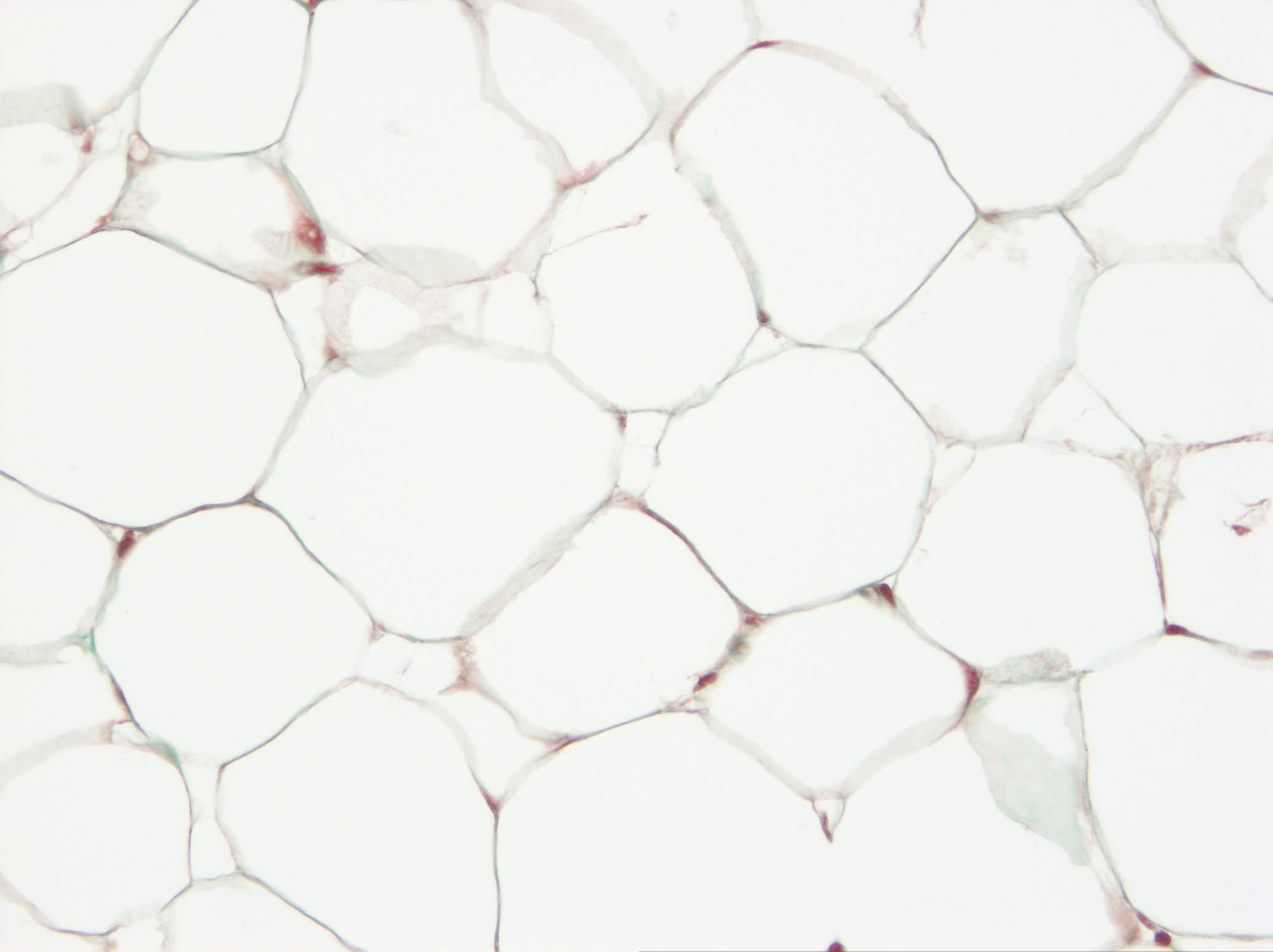
(specific mitochondrial protein of brown adipocytes).This marking enables demonstrating the conversion of white adipocytes into smaller, more easily eliminated brown adipocytes.
After topical application of the product.
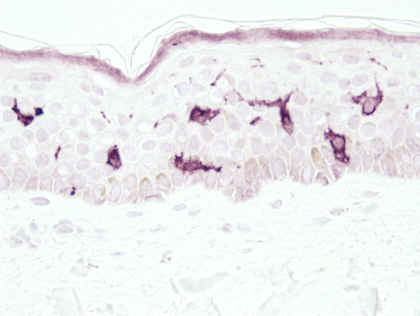
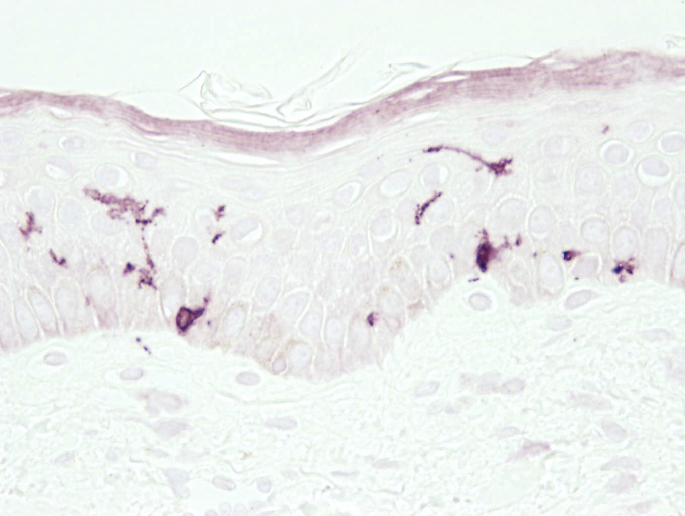
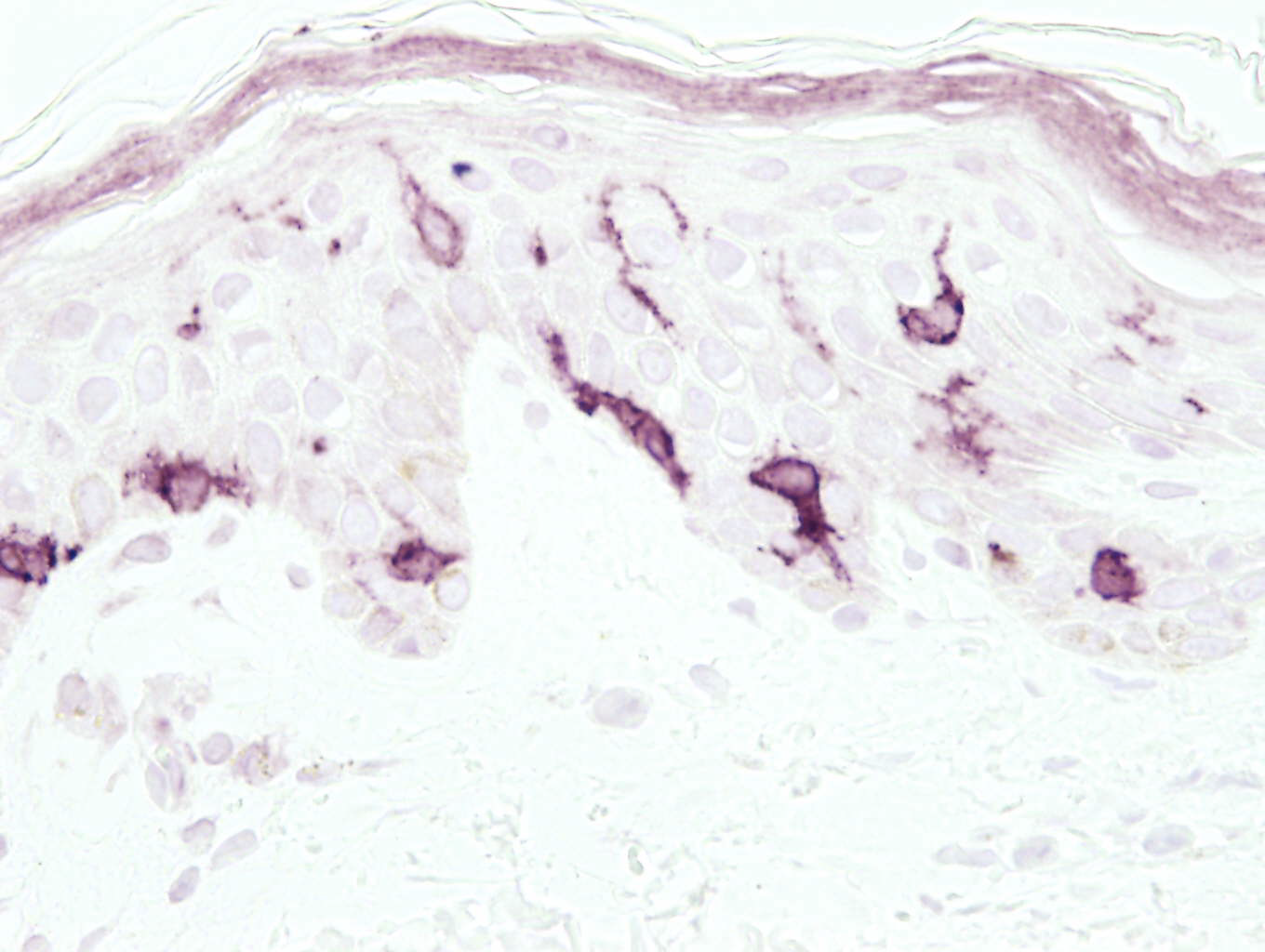
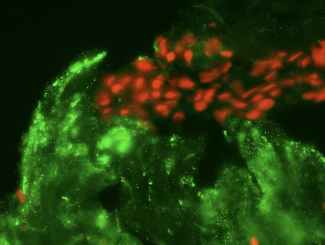
Langerhans cells: CD1a and langerin.

Determination of the irritant character in compliance with GLP (OECD method 439)
Control tolerance
Strong irritant
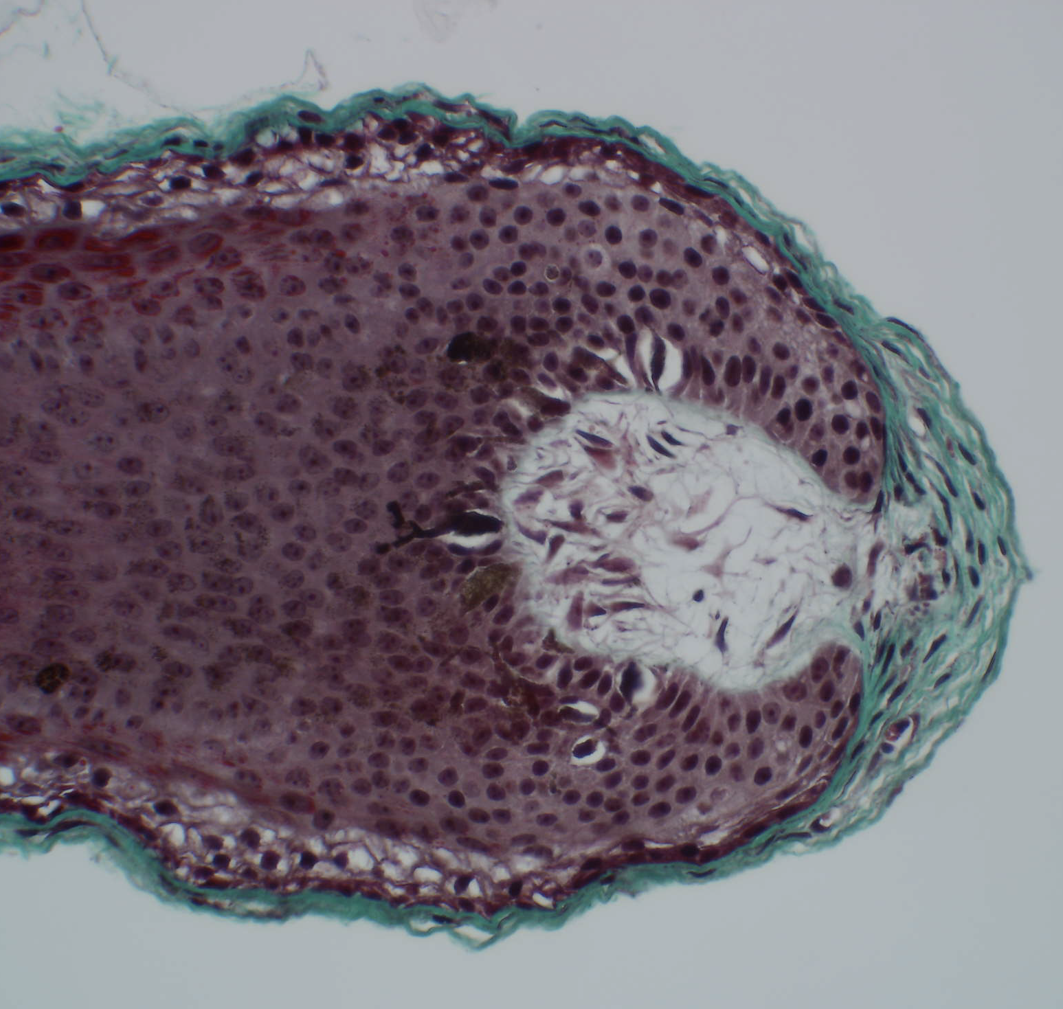
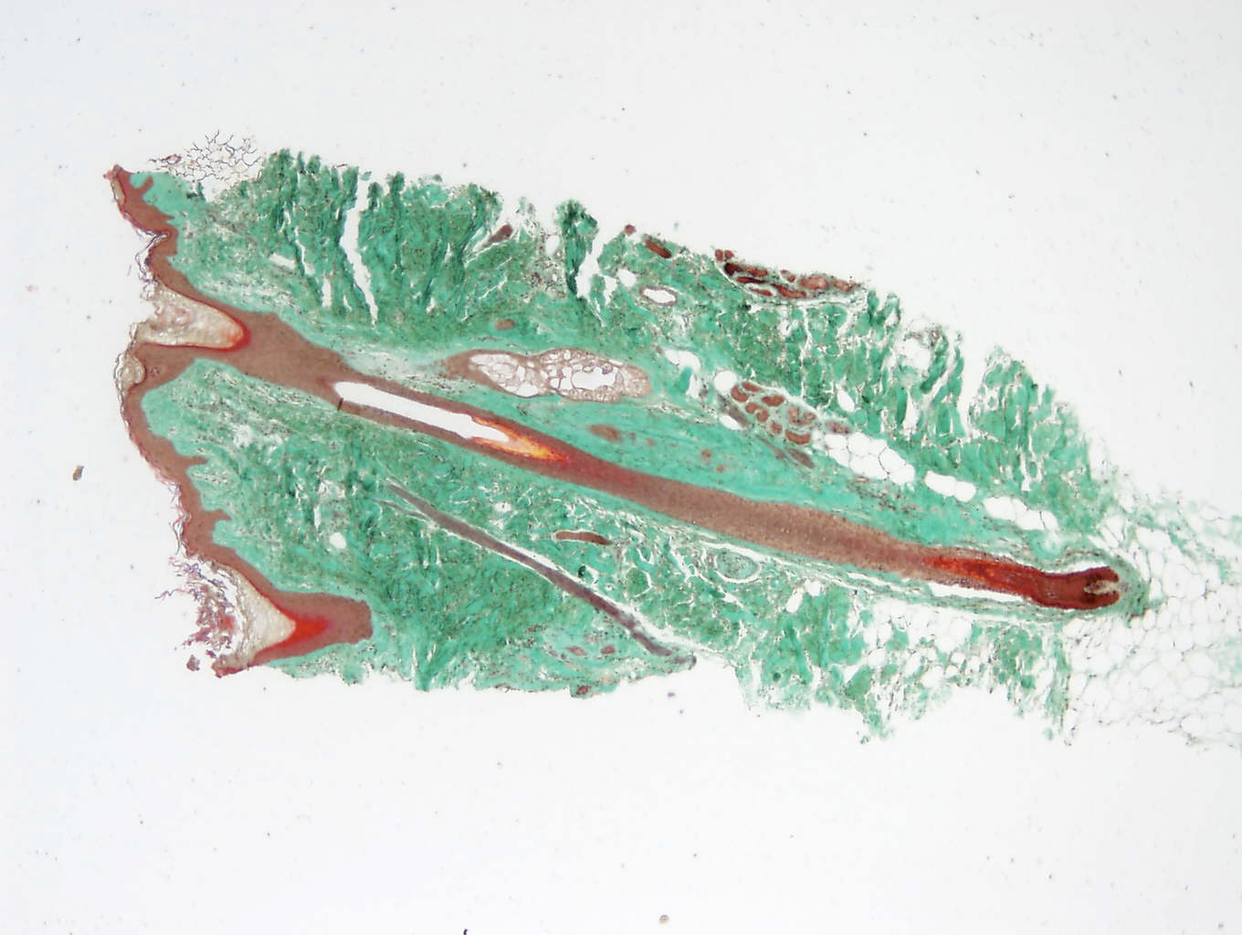
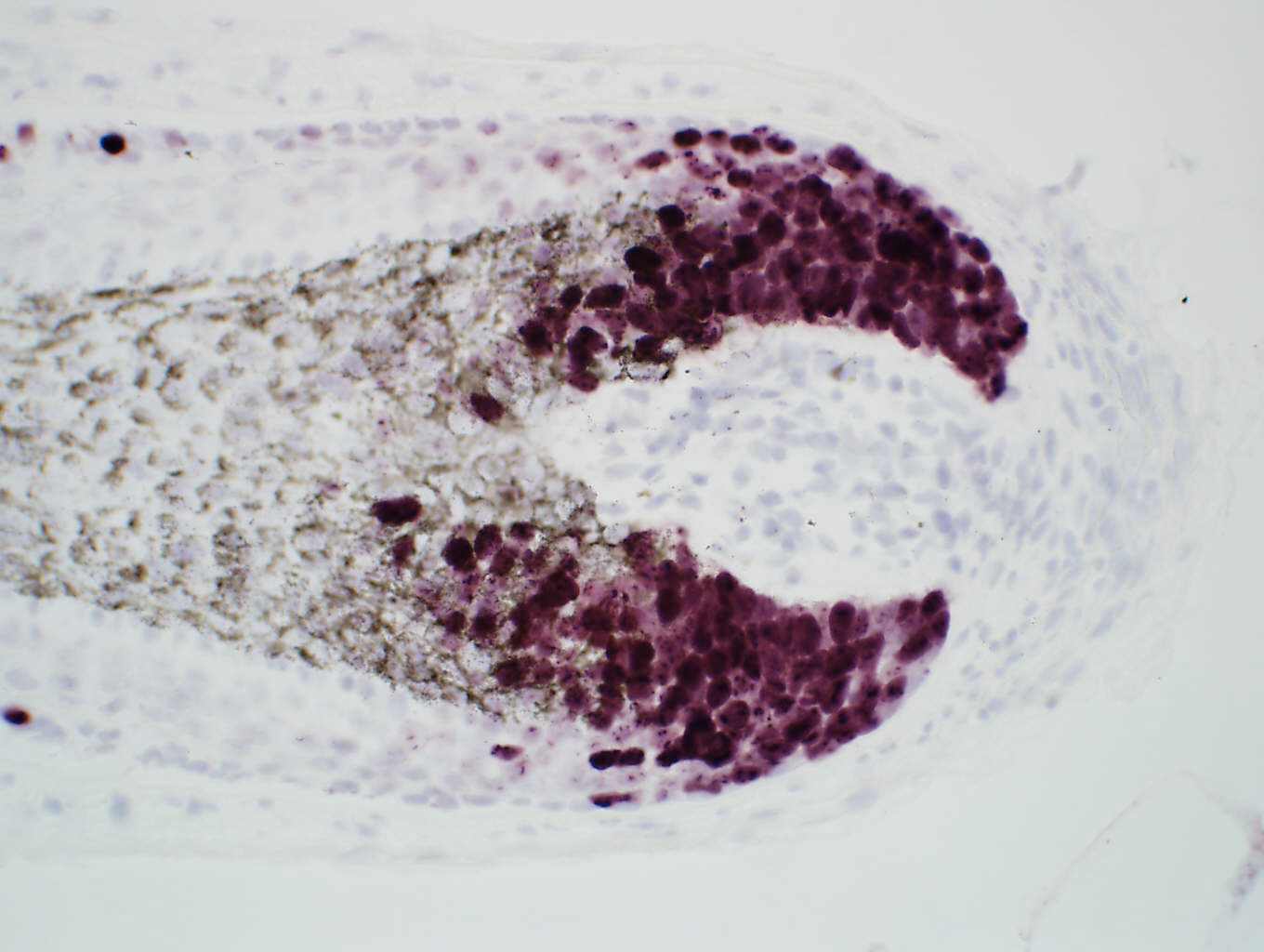
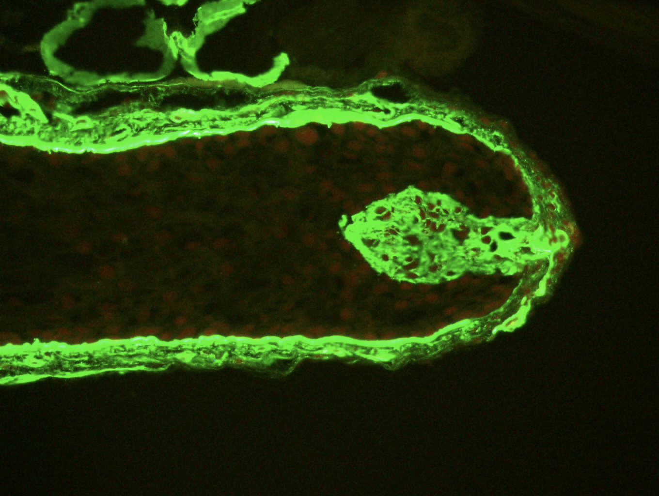
Evaluation of the re-epithelialisation speed (structure of the growth bud).
Cicatrisation bud
Impact of the products on the cutaneous microbiome bacteria (commensal or pathogenic), influence of the product on their adhesion (formation of biofilms, proliferation, etc.).
Process exclusive to BIO-EC Laboratory of exposure of explants to atmospheric pollutants:
– Hydrocarbons,
– Heavy metals,
– Diesel particles,
– Ozone,
– Cigarette smoke, etc…
This device enables pollutants to be vaporised into cloud form and to be put into contact only with the surface of the skin to resemble physiological conditions as closely as possible.
(mitochondrial enzyme)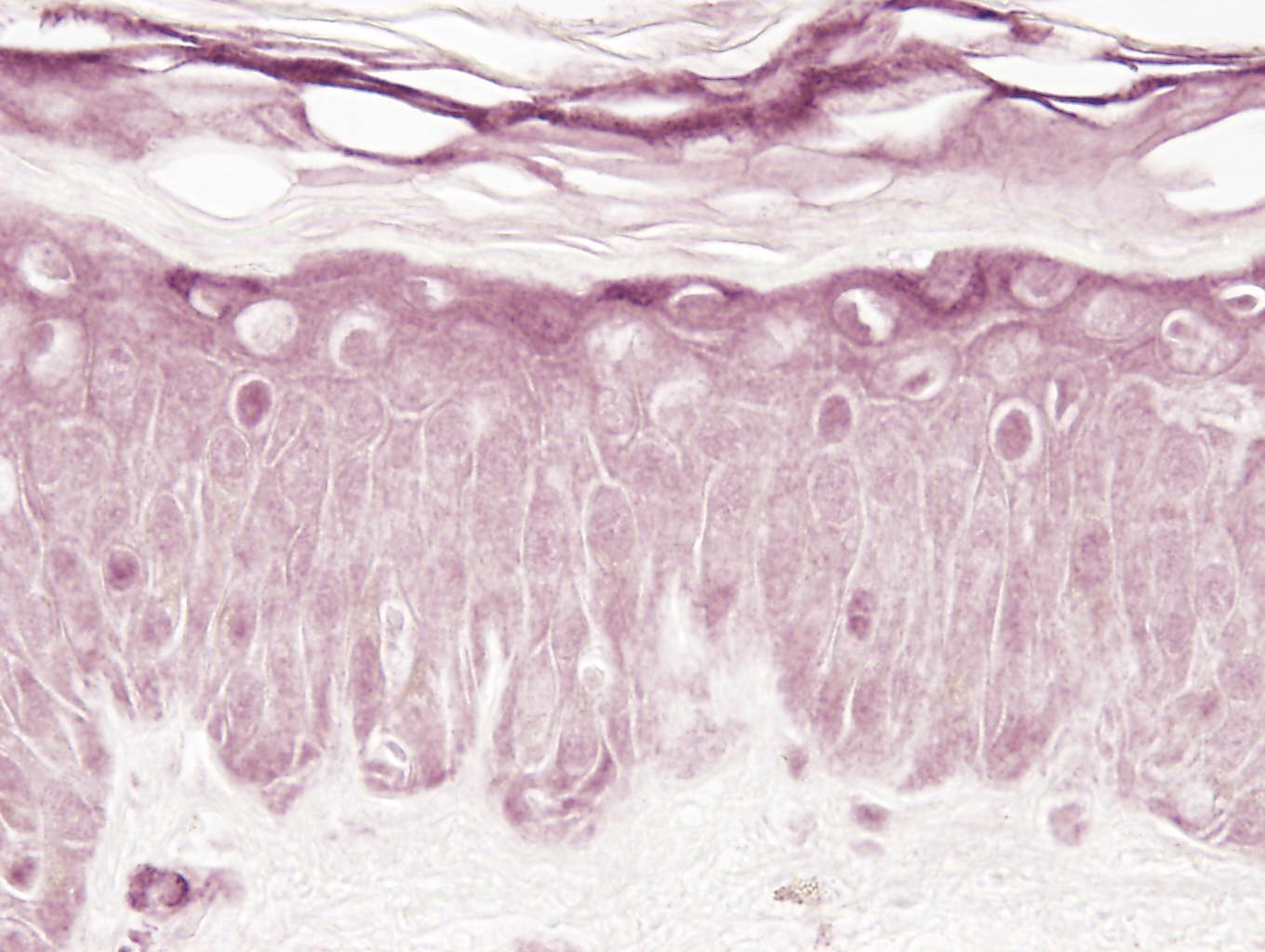
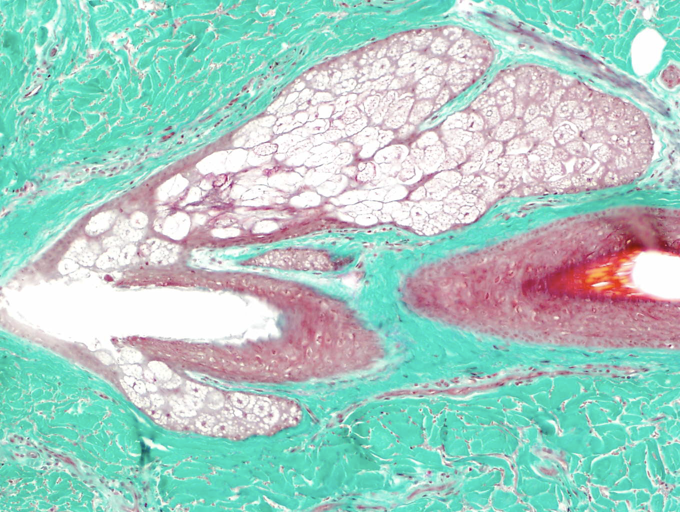
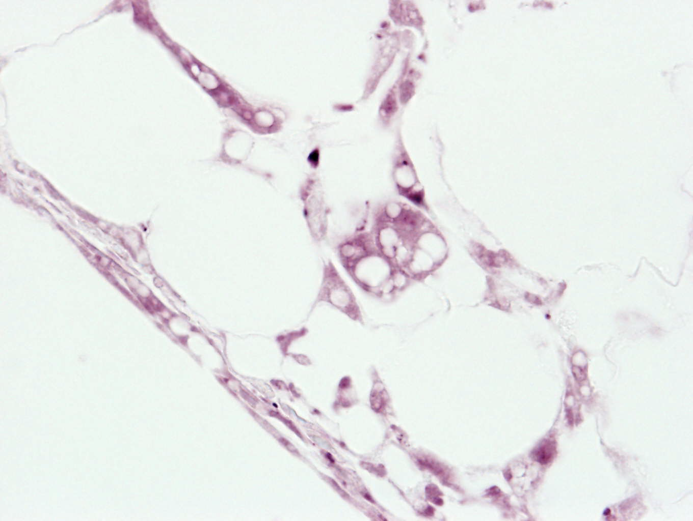
Sebaceous glands
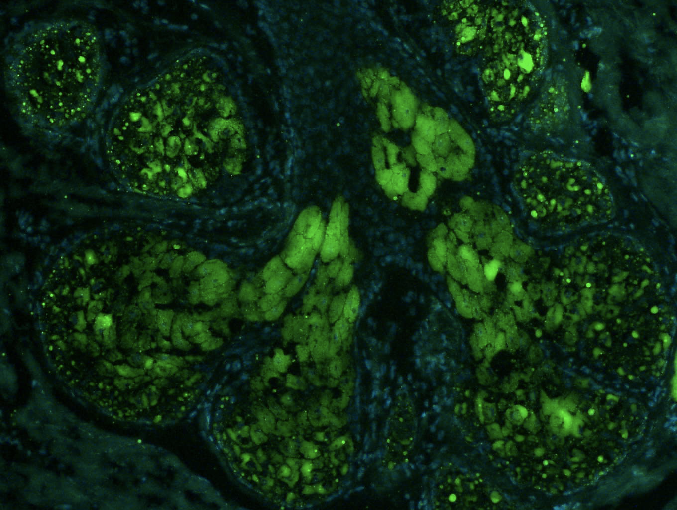
Sebaceous gland sebum, LipidTox
Evaluation after treatment of scalp explants by yeasts involved in the appearance of dandruff.
Whole hair
Entrez votre adresse email pour vous désabonner.
This message only appears when you first log